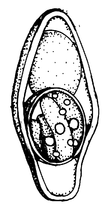
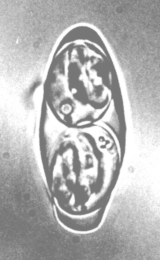
Isospora belli Wenyon, 1923.
Synonyms: Cytospermium hominis Rivolta, 1879
(this name should be regarded
as a nomen nudum); Coccidium isospora Savage and Young, 1917 (this name
should be
regarded as a lapsus calami); Isospora bigemina var. hominis of
Fantham (1917);
Isospora hominis of Fantham (1917); Isospora hominis (Railliet and
Lucet,
1891)
Dobell, 1926, pro parte; Lucetina belli (Wenyon, 1923) Henry and Leblois,
1926.
Type host: Homo sapiens Linnaeus, 1758,
Humans.
Other hosts: Hylobates klossi (Miller,
1903), Gibbon (experimentally, Zamen, 1967).
Type locality: Unknown. During World War I, the
feces of many soldiers were
examined for various reasons; this is when the oocysts of I. belli were first
seen,
but the parasite had not been named at that time.
Geographic distribution: Cosmopolitan, apparently more
common in the tropics
than in the temperate zone.


Description of oocyst: Oocyst shape:
elongate-ellipsoid, constricted at 1
end and asymmetrical at that end; number of walls: 1; wall thickness: 1.1; wall
characteristics: smooth, colorless; L x W: 31.6 x 13.7 (20-33 x 10-19); L/W ratio:
2.3; M: absent; OR: absent; PG: absent. Distinctive features of oocyst: highest L/W
ratio (>2) of any Isospora from primates; tapers at one end, occasionally
asymmetrical at tapered end.
Description of sporocysts and sporozoites:
Sporocyst
shape: subspheroid to
ellipsoid; L x W: 13.6 x 10 (12-15 x 7-11); L/W ratio: 1.4; SB: absent; SSB: absent;
PSB absent; SR: present; SR characteristics: coarse granules scattered throughout
sporocyst; SP: sausage-shaped with 1 acentric RB . Distinctive features of the
sporocyst: small size, lacks SB, 1 acentric RB in SP.
Prevalence: Reasonably uncommon in humans (<1%);
however, Elsdon-Dew and
Freedman (1953) found "many" cases in humans in Natal and reasoned that infections
were often missed because those examining human stools don't look for it. Faust et
al. (1961) listed about 800 reports from the Western Hemisphere, the great majority
from Chile, and Jarpa and Zuloaga (1961) reported 332 cases from Chile. In other
selected reports, Jeffery (1956a, 1958) described I. belli infections in 5
and 32
persons in two schools for "mental defectives" in South Carolina, but in none of
1134 persons in a mental hospital in Georgia (Jeffery, 1956b). Campos et al. (1969)
found 7% of 165 children infected in an orphanage in São Paulo, Brazil, but Briceño
(1952) found it in only three of 46,404 fecal examinations in Venezuela. Kuntz et
al. (1958) found it in the feces of 2% of 50 persons from Istanbul, Turkey, but from
none of 299 persons from Papance, Ankara or Bolu, Turkey.
Sporulation: Exogenous. Oocysts sporulate in 3-5
days at 30-37ºC in 2%
aqueous potassium dichromate solution (Foner, 1939), but may sporulate in 24 hr in
"tropical conditions" (Pellérdy, 1974).
Prepatent period: 9-10 days (?) (Pellérdy,
1974).
Patent period: 16 - 21 days (Pellérdy, 1974); 1-120
(mean 29) days in
patients in two schools for mental defectives in South Carolina (Jeffery, 1956a,
1958).
Site of infection: Enterocytes lining the villi of
the entire small
intestine is where "normal" endogenous development occurs. Lindsay et al. (1997) and
others (Restrepo et al., 1987; Michiels et al., 1994) recently have documented
extraintestinal tissue stages of I. belli in AIDS patients. These tissue cysts may
represent a reservoir of stages that can recolonize the gut to cause recrudescence
of clinical symptoms.
BR>
Material deposited: None.
Cross transmission studies: Zamen (1967) reported
successfully infecting 2
of 3 gibbons with 1-2,000 oocysts of I. belli of human origin. The prepatent
period
in both was 10 days. One gibbon had only a 3 day patent period, the second one was
killed on the second day of patency (11 DPI) to study the tissue stages. Fagot
(1980) mentioned finding "the presence of coccidia" in the feces of a "Gibbon" in a
zoo in Berlin, but no other information was given. Perhaps this also was I.
belli,
but there is no way to know for certain. Other animals such as dog, cat, pig,
domestic rabbit, guinea-pig, mouse, rat and rhesus monkey are refractory to
infection (Foner, 1939; Herrlich and Liebmann, 1943, 1944; Jeffery, 1956a; Robin and
Fondimare, 1960). However, O'Connor, who infected 2 new-born puppies with I.
hominis
(probably I. belli) oocysts, reported that on the 21st day after infection
"weakness
of the hind legs was noticeable, and towards the end of the same day efforts to
maintain the erect position completely failed."
Pathology: In immunocompetent people, symptoms may
include diarrhea,
steatorrhea, headache, fever, malaise, abdominal pain, flatulence, nausea, vomiting,
dehydration and weight loss. However, in immunocompromised individuals (e.g., AIDS
patients), the diarrhea is chronic and highly fluid and can lead to malabsorption
and dehydration that requires hospitalization. It can sometimes be fatal.
Interestingly, I. belli rarely causes diarrhea in AIDS patients in the United
States
(20/14,519, <0.2%), but has a significantly higher prevalence in AIDS patients from
other countries such as Haiti (20/131, 15%) (DeHovitz et al., 1986). It is also
suggested that the infection may be sexually transmitted in homosexual men.
Remarks: For the best reviews on the correct name
of this species see Wenyon
(1923, 1926). There are very few sources that give a complete description of the
sporulated oocysts. Wenyon's (1923) original description only said, "The oocysts
measure from 25 to 30 microns in length by about 12 to 15 microns in breadth."
Reviews (e.g., Levine, 1973; Long and Christie, 1995; Pellérdy, 1974) give
additional mensural data, and clinical reports cite the source and size of their
oocysts only rarely (Jeffery, 1956a). The descriptive parameters given above are
from 25 sporulated oocysts of I. belli, obtained in 1976 from a male
patient at the University of Texas Medical School Hospital, Houston, Texas. They
were maintained in 2.5% aqueous (w/v) potassium dichromate solution and measured and
photographed several weeks after the sample was collected. Both Levine (1973) and
Pellérdy (1974) state that the oocysts sometimes "bear a small micropyle," but we
have never observed this structure in our experience. Levine (1973) also notes, "An
oocyst polar granule may be present in young, incompletely sporulated oocysts, but
quickly disappears." One unique feature of I. belli oocysts is that a certain
percentage of oocysts always seem to develop irregularly producing 1 sporocyst with
8 sporozoites.
Oocysts of I. belli often have been confused with those of
"Isospora"
hominis (i.e., Sarcocystis hominis). Indeed, many reports of I.
hominis actually
were I. belli (e.g., Elsdon-Dew and Freedman, 1953). A number of workers have
thought that I. bell was a synonym of I. hominis, but evidence
presented was not
convincing (also see Wenyon, 1923, 1926). With our new information on the true
nature of "Isospora" hominis (=S. hominis), distinguishing between the
two species
is mandatory. The oocysts of I. belli are not sporulated when passed in the
feces,
while those of S. hominis are and the two species differ considerably in
appearance.
References: Briceño (1952); Campos et al. (1969);
Dobell (1926); DeHovitz et
al. (1986); Elsdon-Dew and Freedman (1953); Faust et al. (1961); Foner (1939);
Herrlich and Liebmann (1943, 1944); Jarpa and Zuloaga (1961); Jeffery (1956a, b,
1958); Kuntz et al. (1958); Levine, (1973); Lindsay et al., (1997); Long and
Christie (1995); Michiels et al. (1994); Pellérdy (1974); Restrepo et al. (1987);
Robin and Fondimare (1960); Wenyon (1923, 1926); Zamen (1967).


