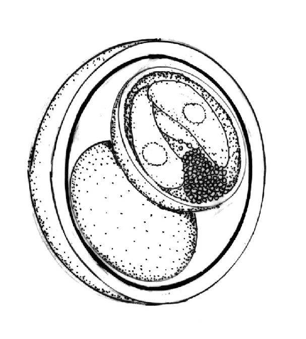
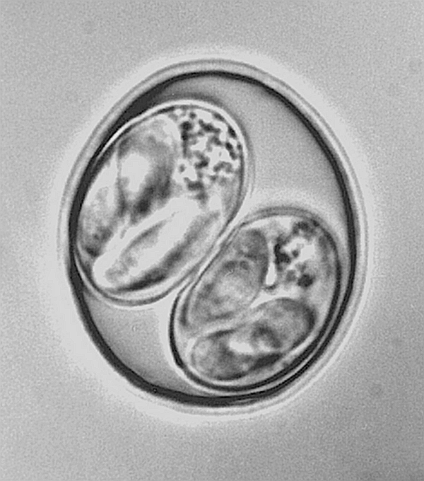
Isospora endocallimici Duszynski and File, 1974.
Type host: Callimico goeldii Thomas, 1904,
Goeldi's marmoset.
Other hosts: None reported to date.
Type locality: Unknown (see Remarks).
Geographic distribution: SOUTH AMERICA: Peru; NORTH
AMERICA: USA: Louisiana, Delta Regional Primate Research Center in Covington.


Description of oocyst: Oocyst shape: ovoid; number
of walls: 2; wall
thickness: 2; wall characteristics: outer layer smooth, ~2/3 of total thickness, and
may taper at pointed end, inner layer appears as a darker line; L x W: 27.8 x 24.0
(25-31 x 21-27); L/W ratio: 1.2 (1.1-1.4); M: absent; OR: absent; PG: absent.
Distinctive features of oocyst: distinctly ovoid shape.
Description of sporocysts and sporozoites:
Sporocyst shape: ellipsoid; L x
W: 17.3 x 12.5 (15-20 x 10-15); L/W ratio: 1.4 (1.2-1.5); SB: absent; SSB: absent;
PSB: absent; SR: present; SR characteristics: granular, usually a compact mass at 1
end of sporocyst; SR L x W: 8 x 5; SP: banana-shaped, 11.3 x 5.4 (10-13 x 5-6), with
sub-central N. Distinctive features of the sporocyst: SP appear sausage-shaped
within sporocyst, but become clearly banana-shaped when they excyst; also, the
compact SR at 1 end of sporocyst.
Prevalence: 5/5 (100 %).
Sporulation: Presumably exogenous. Time and
temperature needed are unknown.
Prepatent and patent periods: Unknown.
Site of infection: Unknown. Oocysts recovered from
feces.
Material deposited: Photoparatype in the USNPC, No.
88513.
Remarks: Five of 5 marmosets housed at the Delta
Regional Primate Research
Center, Tulane University, Covington, Louisiana, U.S.A. were found to be passing
oocysts in their feces from time to time. Two of the hosts were born at the Center
and 3 were imported from Peru so it was not possible to state in which host the
infection originated. Host feces were collected at the Primate Center, but were
examined at The University of New Mexico, Albuquerque, NM. Oocysts were sporulated
upon arrival in Albuquerque. Duszynski and File (1974) described the excystation
process of sporozoites of I. endocallimici and documented the unique folding
of the
sporocyst wall during this process. Speer et al. (1976) describe the ultrastructure
of the sporocyst wall during excystation of sporozoites from their sporocysts.
References: Duszynski and File (1974); Speer et al.
(1976).


