\
Eimeria morainensis Torbett, Marquardt &
Carey, 1982.
Type host: Cynomys gunnisoni
Other hosts: C. leucurus; C.
ludovicianus; Marmota flaviventris; Spermophilus elegans; S.
lateralis; S. richardsonii; S.
townsendii; S. tridecemlineatus; S. variegatus.
Type locality: North America: USA, Colorado Moraine
Park in Rocky Mountain National
Park, TSW, R73W, S29.
Other localities: North America: Canada, Alberta;
USA, Idaho, Utah, Wyoming.


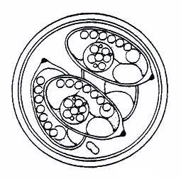
Description of oocyst: Oocyst shape: subspheroid;
wall thickness: 1.5 (from
redescription by Wilber et al., 1994); layers: 2; outer layer proportion of total
thickness: not given; outer layer colour: gray to blue-gray; outer layer texture:
smooth; inner wall characteristics: smooth, colourless; micropyle: absent; OR:
absent; PG: present; number of PGs: 1; PG shape: often bilobed (Wilber et al.,
1994); PG L x W: not given; size: 20.3 x 19.8 (19-26 x 18-21); L/W ratio: 1.1
(1.0-1.1). Distinctive features of oocyst: none.
Description of sporocysts and sporozoites:
Sporocyst
shape: ellipsoid; size: 12.1 x
6.9 (9-14 x 6-9); SB: present; SB L x W: not given; SB characteristics: prominent,
dark, button-like (Wilber et al, 1994); SSB: absent; PSB: absent; SR: present; SR
characteristics: compact mass off centre or in centre of sporocyst with size and
amount varying, appears to be membrane-bound (in drawing); 2nd SR a row of globules,
usually along the side of sporocyst (Wilber et al., 1994); SR size: not given; SP:
elongated with refractile bodies at opposite ends. Distinctive features of
sporocyst: 2 different SR.
Material deposited: USNPC No. 82933
(phototype).
Remarks: Eimeria morainensis was described
in S. lateralis by Torbett et al.,
(1982). McAllister et al. (1991) reported it from S. mexicanus, but Wilber et
al.
(1994) suggested the oocysts seen by McAllister et al. (1991) more closely resembled
E. adaensis (= E. vilasi, see below). Thomas & Stanton (1994) found
E. morainensis
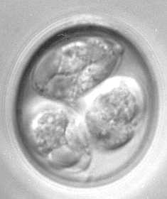
in M. flaviventris (Thomas, unpubl. data). Wilber et al. (1994) redescribed
E.
morainensis, noting the presence of a second sporocyst residuum which appears in
the
photomicrographs, but not in the original description or drawing of Torbett et al.,
(1982).
References: McAllister et al. (1991); Seville
(1997); Seville et al. (1992); Seville
& Stanton (1993b); Shults et al. (1990); Stanton et al. (1992); Thomas & Stanton
(1994); Torbett et al. (1982); Wilber et al. (1994).


