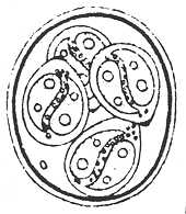
Eimeria suncus Ahluwalia, Singh, Arora, Mandal and Sarkar, 1979
Type host: Suncus murinus murinus (Linnaeus, 1766), House shrew.
Other hosts: S. m. soccatus (Hodgson).
Type locality: ASIA: India: West Bengal, Mathura Veterinary College Campus.
Geographic distribution: ASIA: India.

Description of oocyst: Oocyst shape: subspheroid;
number of walls: 2;
wall thickness: 1.7-1.9;
wall characteristics: outer, smooth, yellow, thinner than inner layer;
L x W: 19.5 x 15.2 (18-22 x 15.5-17);
L/W ratio: 1.3;
M: absent;
OR: absent;
PG: reported as absent (?).
Distinctive features of oocyst: the authors state, "a clear micropylar cap is visible on the wall of the oocyst,
but no ditinct micropyle is seen in any of the oocysts examined." However, they did not include a micropyle
cap in their line drawing, so our conclusion is that the PG was confused for a micropylar cap.
Description of sporocysts and sporozoites:
Sporocyst shape: lemon-shaped;
L x W: 10.5 x 7.8 (9.5-11 x 6.5-9);
L/W ratio: 1.35;
SB: present;
SSB: absent;
PSB: absent;
SR: present;
SR characteristics: beaded-like globular mass, scattered;
SP: comma- to banana-shaped, 4.5 x 3.5 (4-5.5 x 3-4), with a round RB at each end of sporocyst and SPs with 2 RB.
Distinctive features of sporocyst: nipple-like SB at thickened end of sporocyst and SPs with 2 RB.
Prevalence: 12/~100 (12%) in the type host collected from 2 localities
in West Bengal; 1/11 (9%) of S. m. m. soccatus "collected...from various parts of the country (i.e., India)."
Sporulation: Exogenous. Oocysts sporulated in 48 hours in 2.5% potassium dichromate solution at 20-22 C.
Prepatent and patent periods: Unknown.
Site of infection: "Columnar epithelial cells in sections of parts of the small intestine" Ahluwalia et al. (1979).
Endogenous development: Bi- and multinucleate meronts were seen mostly in the subepithelium
of the small intestine. Trophozoites were 4.9 x 4.1 (4-6 x 3-5) with a nucleus that was 3.3 x 2.1 (3-4 x 1.5-2.3).
Binucleate meronts were 3.9 x 2.9 (2.8-5 x 2-4) and multinucleate meronts, with 12 merozoites, were 6.2 x 4.8 (4-9 x 3-6).
Gametocytes also were found in the subepithelium, sometimes just above the muscularis mucosa. Young
microgametocytes, with numerous scattered nuclei, were 4.9 x 4.0 (4.5-5 x 3.8-4.2). Mature microgamonts, with comma-shaped
microgametes arranged peripherally, were 8.0 x 5.9 (7-8.3 x 5.7-6). Young macrogametocytes were 5 x 4 (4-6 x 3-4.5) with
a nucleus ~1.5. Mature macrogamonts, with a centrally-located nucleus (~2) and vacuolated cytoplasm, measured 6.3 x 4.3 (5-7 c 3.5-5).
Zygotes were somewhat larger, 7.2 x 6.0 (6-8 x 5-7).
Materials deposited: None. Ahluwalia et al. (1979) said, "Holotype: Z.S.I. Registration No."
but did not give the number and Bandyopadhyay and Das Gupta (1984), who made tissue sections from an
infected host, did not deposit them anywhere.
Remarks: Ahluwalia et al. (1979) stated that, "stained sections exhibit
oocysts, macrogametocytes and microgametocytes inside coulmnar epithelial cells," but gave no drawings or
photomicrographs; however, Bandyopadhyay and Das Gupta (1984) described and measured the endogenous
stages from 1 S. m. soccatus they found in Darjeeling, West Bengal. The oocysts they described
from fecal material were quite similar to those first described by Ahluwalia et al. (1979): subspherical oocysts
were 19.6 x 18 (17-21 x 16-19.5) with a smooth, 2-layered wall ~1.2-1.5; outer layer thinner than inner
layer; OR absent, ovoid sporocysts were 10.2 x 6.8 (9-11 x 6-7.5) with a SB and a SR of scattered, fine
globules. Bandyopadhyay and Das Gupta (1984) repeated the same error of Ahluwalia et al. (1979), mistaking a PG
for a micropyle.
References: Ahluwalia et al. (1979); Bandyopadhyay and Das Gupta (1984).

